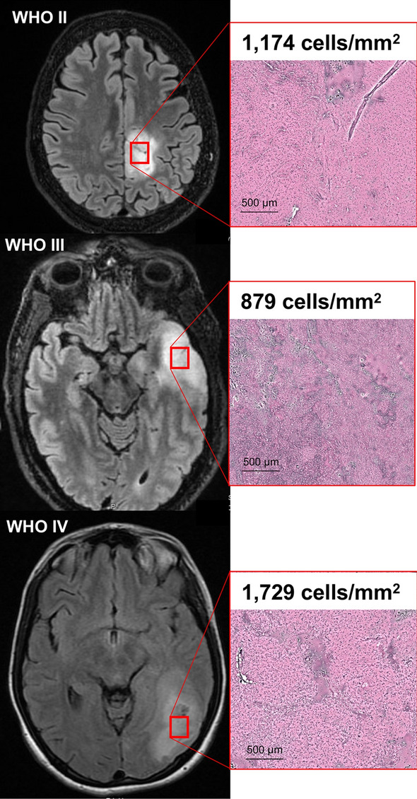
Figure 1.

T2-weighted fluid attenuation inversion recovery (FLAIR) images of three participants with World Health Organization (WHO) grade II, III, and IV glioma (left). Red boxes indicate regions of FLAIR signal abnormality biopsied for pathologic examination and cell-counting with Raman scattering microscopy (right). Pseudo-H&E images of fresh biopsy specimens demonstrate ablation of normal cytoarchitecture across all WHO tumor grades. Here, lesion cellularity and necrotic features escalate with increasing WHO grade.
