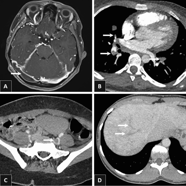
Figure 2.
Various examples of eosinophil-related venous thrombosis. Right transverse sinus thrombosis on brain Magnetic Resonance Imaging (A), bilateral pulmonary embolism (middle lobe medial segment, right posterior basal segment left anterior basal segment) on Computed Tomography pulmonary angiography (B), left common iliac vein thrombosis on Computed Tomography venography (C), right and middle hepatic thromboses on contrast-enhanced abdominal Computed Tomography at late portal phase (D).

