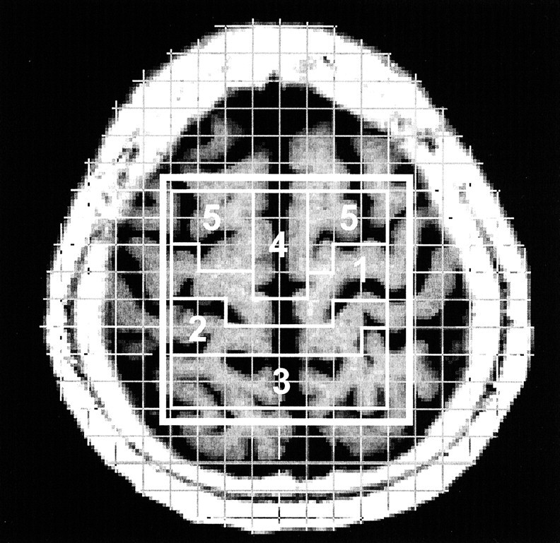
Fig 1.

Axial T1-weighted MR image with a superimposed phase-encoding grid and volumes of interest. Voxels selected for the five cortical-subcortical regions are outlined: 1 indicates the precentral gyrus; 2, the postcentral gyrus; 3, the superior parietal lobule; 4, the supplementary motor area; and 5, the premotor cortex.
