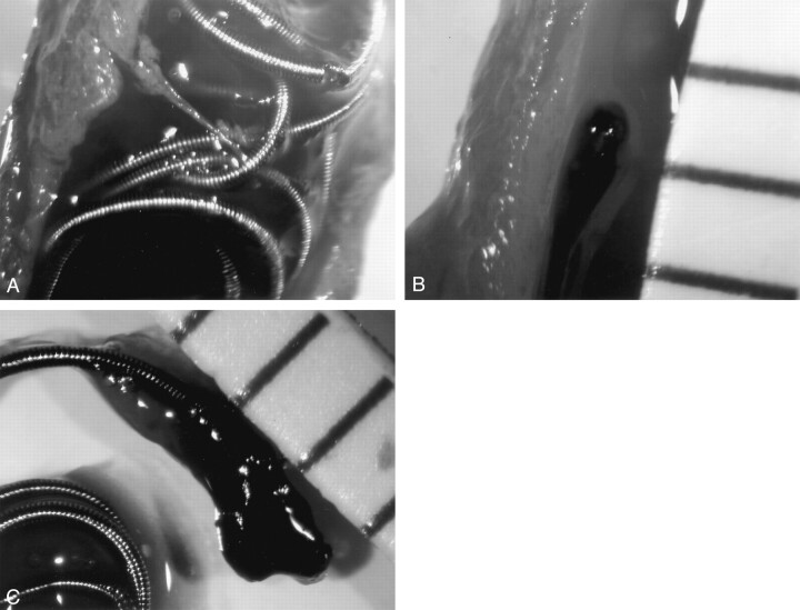
Fig 4.
Depiction of the detachment process of a conventional coil (GDC-18, 8 mm × 20 cm) in the canine carotid artery.
A, Immediately after detachment of the coil, the segment of the artery is ligated and opened by use of a longitudinal arteriotomy. Without arteriotomy, multiple and tiny gas bubbles surround the coil.
B, A thrombus with elongated appearance is seen at the proximal tail of the detached coil.
C, After removal of the coil from the artery, the thrombus measures 3 mm in diameter.

