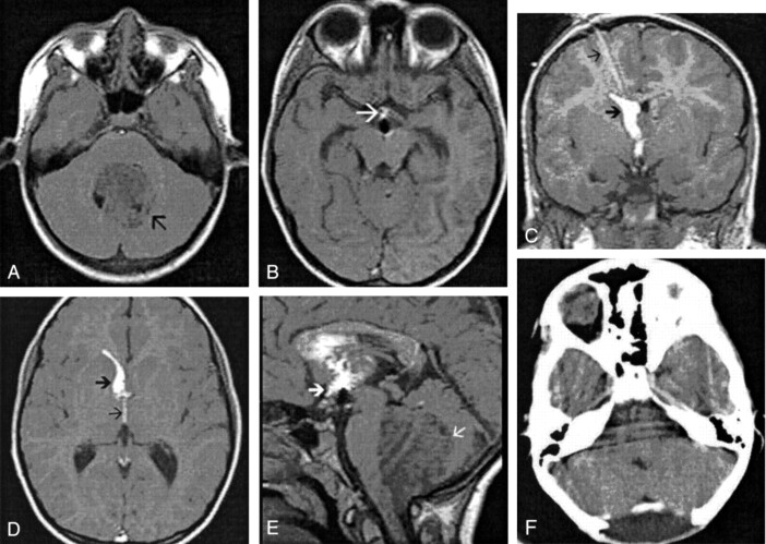
Fig 4.
8-year-old boy with a medulloblastoma and hydrocephalus who underwent an emergency third ventriculostomy before excision of the tumor.
A, Nonenhanced axial T1-weighted MR image shows a large hypointense tumor (arrow) in the posterior fossa.
B, Axial MR ventriculogram after third ventriculostomy shows contrast material in the anteroinferior third ventricle (arrow). Note that there is no contrast material in the adjacent suprasellar and sylvian cisterns.
C, Coronal ventriculogram after third ventriculostomy shows contrast material along the external ventricular drain (thin arrow), the frontal horn of the right lateral ventricle (thick arrow), and the third ventricle. Note that no contrast material is seen in the suprasellar cisterns.
D, Axial MR ventriculogram after third ventriculostomy shows contrast material in the right frontal horn (thick arrow) and third ventricle (thin arrow).
E, Sagittal MR ventriculogram after third ventriculostomy shows contrast material in the dependent portion of the lateral ventricle and the third ventricle (thick arrow). There is no contrast material seen in suprasellar cisterns. Note the large hypointense tumor (thin arrow) in the posterior fossa.
F, Postoperative contrast-enhanced CT scan through the posterior fossa shows complete tumor excision.

