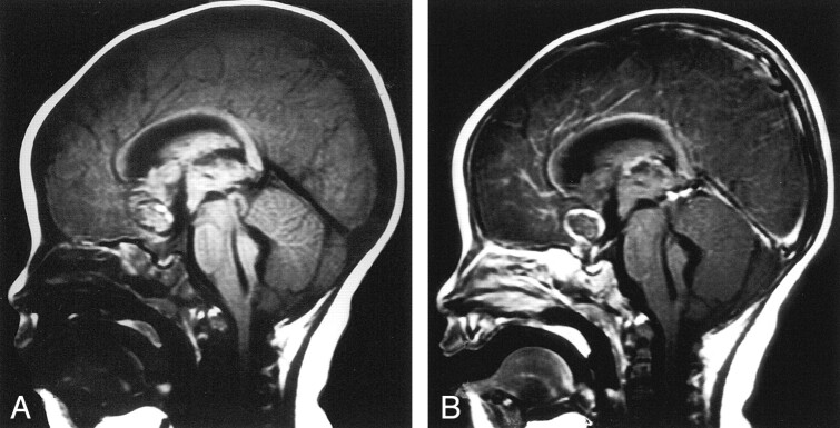
Fig 3.
Sagittal MR images.
A, Nonenhanced T1-weighted image shows hyperacute hemorrhage in the frontal lobe parenchyma extending into the ventricular system. A hypointense ring with a partial, hyperintense inner margin was noted at the center of the parenchymal hematoma.
B, Contrast-enhanced image shows enhancement of the outer wall of an aneurysm of the anterior communicating artery.

