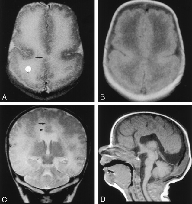
Fig 1.
MR images obtained when the patient was aged 3 weeks.
A and B, Fast spin-echo T2-weighted (TR/TE/NEX, 5460/106/2) (A) and spin-echo T1-weighted (540/14/1) (B) images show interhemispheric fusion of the gray matter and white matter (arrow in A) and an absence of the IHF in the posterior frontal and parietal lobes. The gyral pattern is abnormal, showing an increased cortical thickness and extensive irregularity of the cortical–white matter junction in the bilateral frontal and parietal lobes.
C, Coronal fast spin-echo T2-weighted image shows interhemispheric fusion of the gray and white matter (arrow) and subcortical heterotopia (arrowhead).
D, Sagittal spin-echo T1-weighted image shows a hypoplastic basis pontis.

