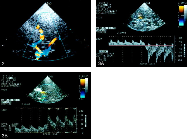
Fig 3.
Doppler spectra.
A, Spectrum shows blood flow reversal in the A1 during ipsilateral carotid compression indicating functional patency of the AcoA.
B, Spectrum shows blood flow velocity increase of more than 20% in the P1 during ipsilateral carotid compression indicating functional patency of the PcoA.

