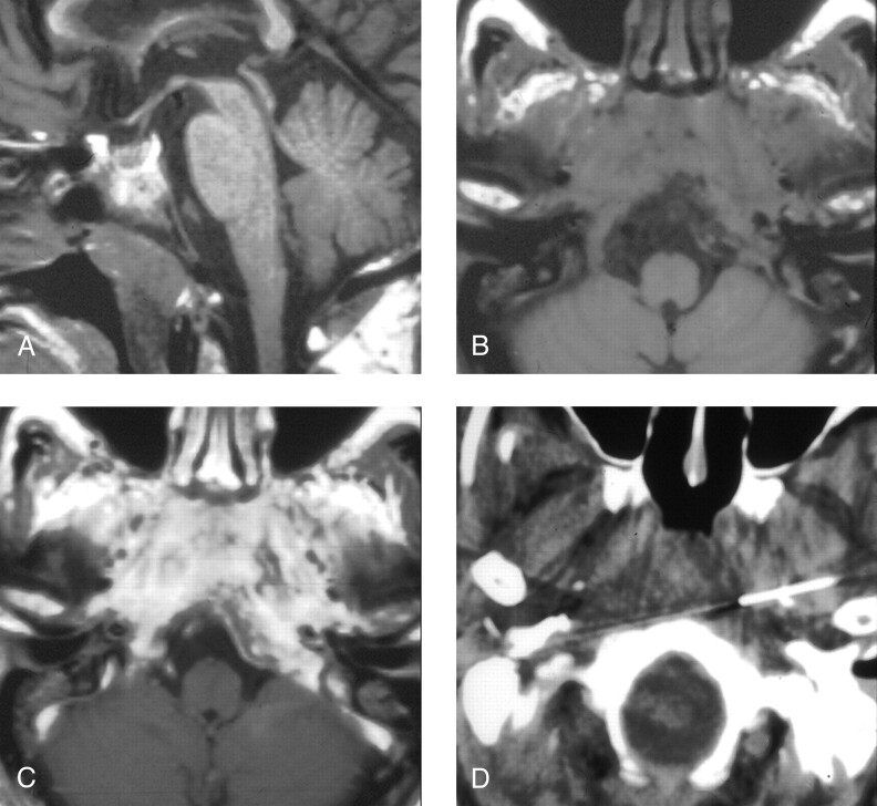
Fig 3.
Patient 4, a 72-year-old man presenting with headache, dysphagia, and progressive hoarseness.
A, Sagittal T1-weighted image (600/11/2) demonstrates abnormally hypointense signal in the lower clivus.
B, Axial T1-weighted image (600/11/2) demonstrates abnormal soft tissue isointense to muscle infiltrating submucosally into the nasopharyngeal tissues, around the carotid arteries, and back to abut the abnormal clivus.
C, Contrast-enhanced axial T1-weighted image (600/11/2) demonstrates marked enhancement within the infiltrated tissue.
D, CT-guided FNA of the preclival soft tissues shows the needle tips anterior to the lower clivus.

