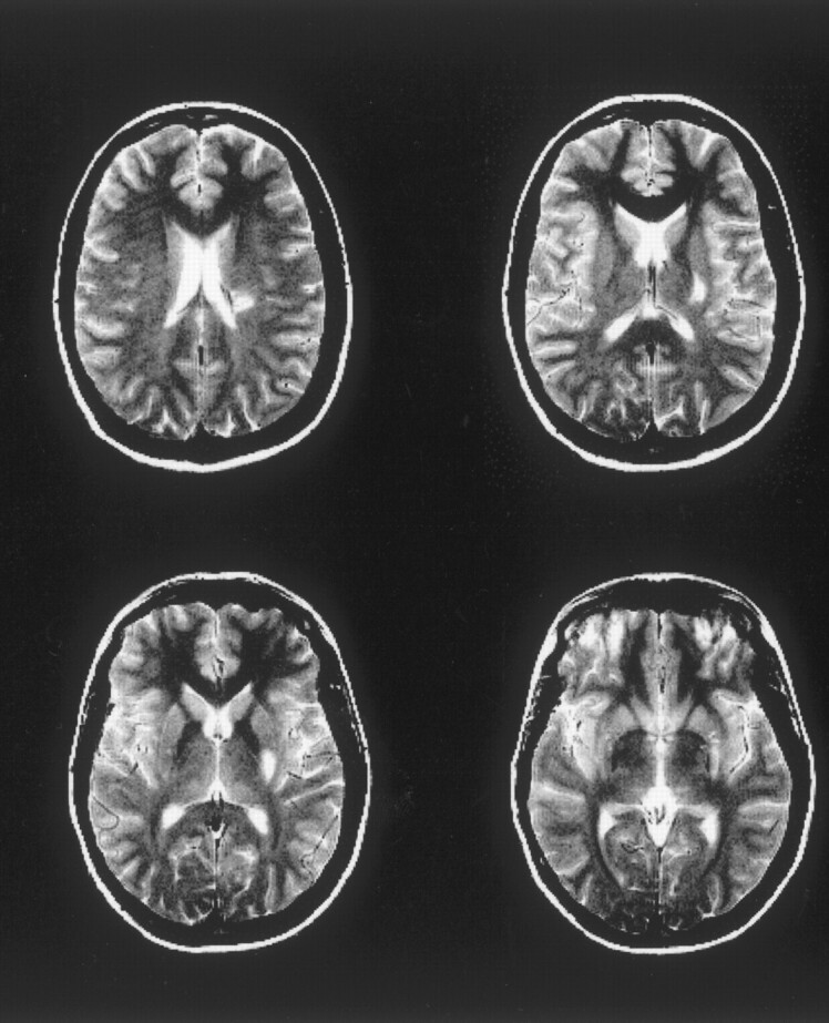
Fig 1.

Patient 1. Whole lesion, as assessed by T2-weighted imaging at the third examination, 392 hours after the onset of symptoms. This demonstrates the typical lesion location in our patients, in all cases affecting the posterior limb of the internal capsule and the medial globus pallidus. In addition, the corona radiata is involved.
