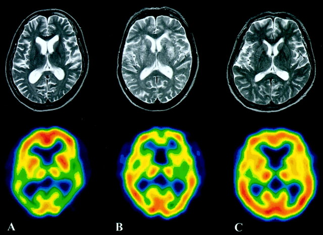
Fig 2.
A representative case from each group (same cases as in Fig 1). Upper images are T2-weighted MR images; lower images are SPECT images.
A, A 69-year-old woman with AD. T2-weighted MR image does not show any clear abnormality. SPECT image shows blood flow reduction in the bilateral temporoparietal lobes.
B, A 67-year-old man with VaD. Multiple small infarcts are observed in the bilateral basal ganglia on the T2-weighted MR image. A frontal region decrease of CBF is noted on the SPECT image.
C, A 70-year-old female age-matched control subject.

