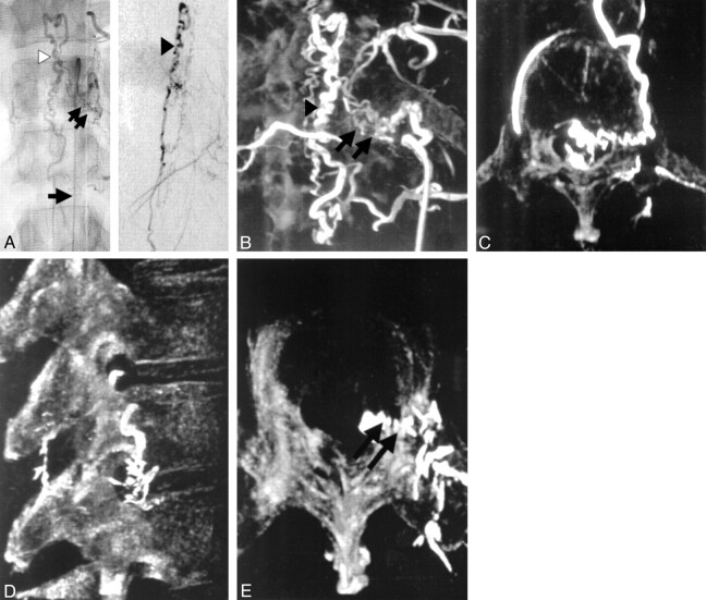
Fig 1.
Patient 8, 54-year-old male patient with spinal dural AVF.
A, Unsubtracted anteroposterior (AP) (left) and subtracted lateral (right) spinal angiograms of left T12 intercostal artery show the spinal dural fistula. (arrow [AP projection], angiographic catheter; double arrow [AP projection], site of fistula at the dural sleeve; arrowhead [AP and lateral projection], anterior draining spinal vein).
B, 3D reconstruction of T12 intercostal rotational injection with partial opacification of the spinal column shows the site of the fistula (double arrow) in relation to the venous drainage (arrowhead).
C, Computer-generated rotation of the reconstructed image with a thin region of interest allows for a better demonstration of the fistula entering the intervertebral foramen.
D, Lateral projection of the 3D-reconstructed rotational image after successful obliteration of the fistula.
E, Similar view as in C, with the glue cast seen entering the spinal canal via the intervertebral foramen (double arrow).

