Fig 2.
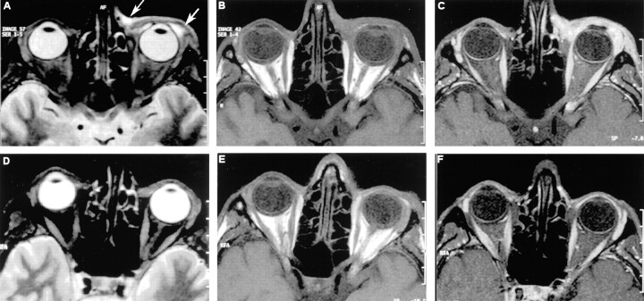
MR images obtained a 38-year-old man who developed AIDS-related Kaposi sarcoma of the conjunctiva and lacrimal gland. All axial sections were tilted to the level of the optic nerve; coronal sections were perpendicular to axial sections. Posttreatment images were obtained 3 months after pretreatment images.
A, Axial turbo inversion recovery magnitude (4000/30 [TR/TE]) MR image of the orbita shows an abnormal mass (arrows) with high signal intensity that involves the conjunctiva and lacrimal gland.
B, Axial T1-weighted (735/12) MR image of the orbita shows an abnormal mass that is isointense relative to muscular tissue (arrows) and involves the conjunctiva and lacrimal gland.
C, Corresponding postcontrast T1-weighted (735/12) MR image shows a marked enhancement of the abnormal tissue of the lower eyelid.
D, Corresponding posttreatment axial turbo inversion recovery magnitude (4000/30).
E, Corresponding posttreatment axial T1-weighted (735/12).
F, Corresponding posttreatment postcontrast T1-weighted (735/12)

