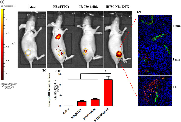
Figure 10.
Tumor-specific targeting and NIRF imaging of IR780-NBs-DTX in subcutaneous xenotransplanted pancreatic cancer in vivo. (a) Nude mice bearing tumors derived from the Mia-Paca2 cell line were detected via the IVIS Lumina II system at 1 h after being injected with different contrast agents. The yellow dotted lines represent the tumor outline. (b) Comparison of the average fluorescence intensity of tumors in different groups. *P < 0.05, significantly different from the average fluorescence intensity of tumors in the saline group, NBs (FITC) group and IR-780 iodide group. (c) Immunofluorescence detection of tumor tissues for the IR780-NBs-DTX” group at 1 min, 5 min and 1 h after injection of contrast agents through the caudal vein.

