Figure 3.
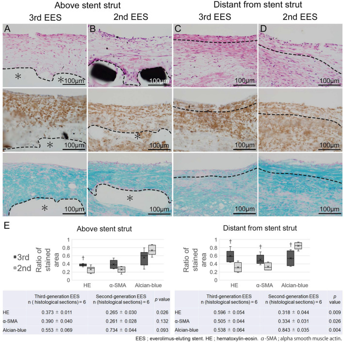
Neointimal tissue characteristics of the 4-week stent implantation group of low-density lipoprotein receptor knockout minipigs. (A–D) High-power magnification images of the neointima from the 4-week group of coronary arteries of low-density lipoprotein receptor knockout (LDLR −/−) minipigs implanted with the third-generation abluminal biodegradable-polymer everolimus-eluting stents (3rd EES, A, C) and second-generation durable-polymer everolimus-eluting stents (2nd EES, B, D) (upper panel: Hematoxylin–eosin, middle panel: alpha-Smooth muscle actin, lower panel; acian-blue). (A, B) Show the neointima above stent struts and (C, D) show the neointima distant from the stent struts. The part above the dotted line corresponds to the neointima and the part below to the intima and media. (A, C) Neointima after the 3rd EES shows several compact layers of tightly arranged smooth muscle cells (SMCs), close to the luminal surface. (B, D) Neointima after the 2nd EES. Although thicker than that seen after the 3rd EES implantation, the neointima consists of proteoglycan-rich tissue and its density of SMCs is relatively low. (E) Neointima above the stent struts of the 3rd EES demonstrated significantly higher SMC density by hematoxylin–eosin staining than those with the 2nd EES. Neointima distant from the stent struts of the 3rd EES shows statistically significant differences with higher SMC and a lower density of alcian-blue-positive proteoglycans. †p < 0.05. (A, B *; stent struts).

