
fig 1.
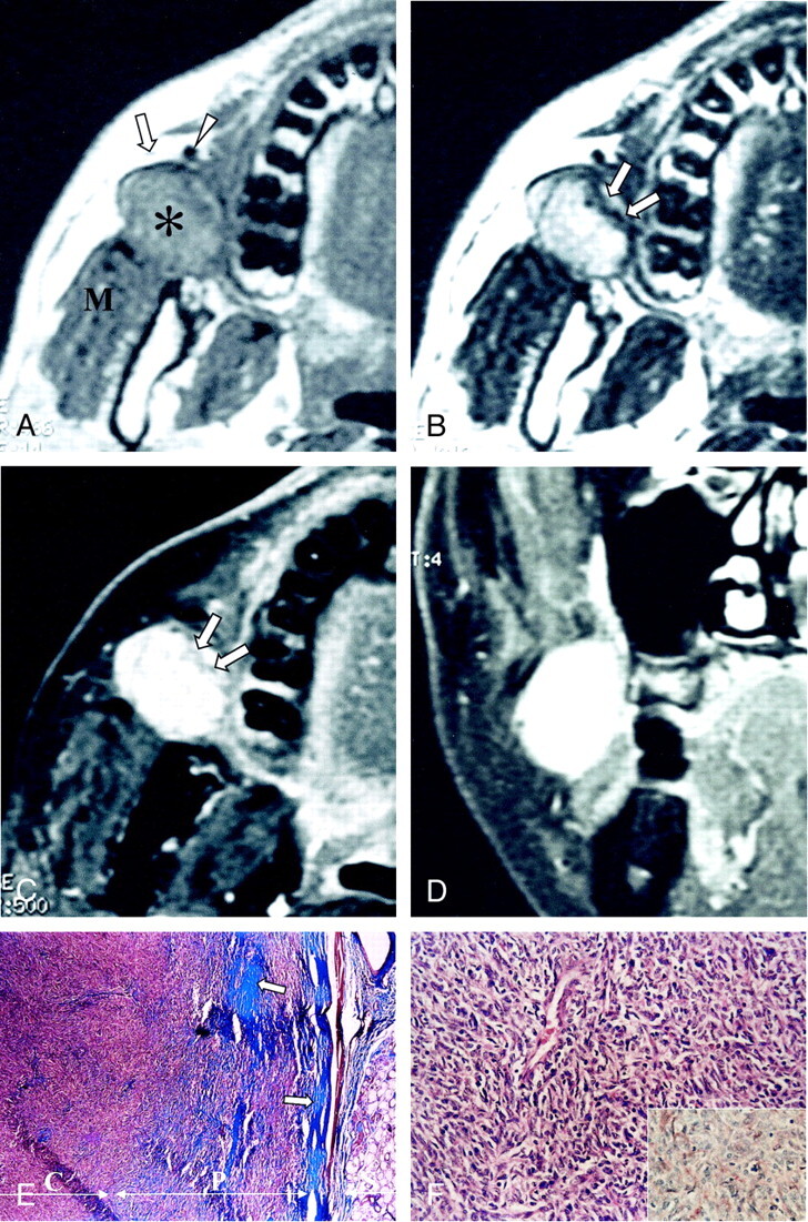
Images in a 46-year-old man with a painless right cheek mass.
A, Axial T1-weighed MR image (466/14 [TR/TE]) shows a well-marginated mass (asterisk) that is isointense to the muscle in the right posterior buccal space. The mass displaces the parotid duct (arrow) and the facial vein (arrowhead) anteriorly and compresses the masseter muscle (M) posteriorly.
B, Axial T2-weighted MR image (4016/104) shows that the mass has mainly high signal intensity, with linear signal isointensity in the medial peripheral portion (arrows).
C and D, Axial (C) and coronal (D) contrast-enhanced T1-weighted MR images (500/14) reveal homogeneously strong enhancement of the mass, with the less enhanced portion in the medial peripheral portion (arrows in C); this portion corresponds to the hypocellular collagenous sclerotic area found at pathologic correlation.
E, Low-power photomicrograph shows a well-circumscribed mass adjacent to a minor salivary gland in the buccal wall. The peripheral portion (P) is hypocellular but more collagenous (blue, arrows) compared with the central portion (C); this finding correlates with the difference in signal intensity between the central and peripheral portions on the T2-weighted and contrast-enhanced T1-weighted images (Masson-trichrome, magnification × 40).
F, Photomicrograph shows that the tumor is composed of mostly a hypercellular proliferation of spindle cells arranged haphazardly in a collagenous background. The spindle cells show elongated bland nuclei surrounded by a rim of amphophilic cytoplasm (hematoxylin-eosin, magnification × 200). The inset shows that the tumor cells and capillary endothelial cells have immunohistochemically positive CD-34 results; this finding is consistent with that of a solitary fibrous tumor (avidin-biotin conjugation, magnification × 200).