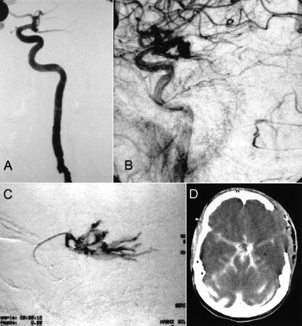
fig 1.

Case 1.
A, Lateral projection arteriogram of the left internal carotid artery reveals a posterior communicating artery aneurysm.
B, Lateral projection arteriogram of the left internal carotid artery, obtained immediately after aneurysm rupture that occurred after placement of the first coil, shows the rupture and massive extravasation of contrast material.
C, Selective angiogram obtained via the microcatheter.
D, Axial view CT scan obtained after clip placement in the aneurysm shows massive brain edema and contrast agent in the subarachnoid space.
