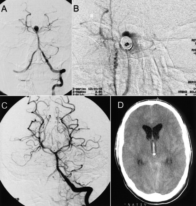
fig 2.

Case 2.
A, Frontal projection arteriogram of the left vertebral artery shows a basilar tip aneurysm.
B, Frontal projection arteriogram of the right vertebral artery, obtained after placement of two GDCs, reveals slight extravasation of contrast material (magnification).
C, Frontal projection arteriogram of the left vertebral artery shows complete embolization of the basilar artery tip aneurysm.
D, Axial view CT scan depicts slight extravasation of contrast agent and blood.
