
fig 3.
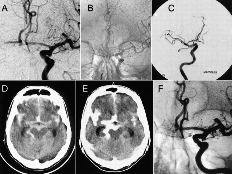
Case 3.
A, Frontal projection arteriogram of the right internal carotid artery shows a small, caudally oriented anterior communicating artery aneurysm.
B and C, Arteriograms of the right internal carotid artery, obtained after placement of one GDC, show that the coil partially protrudes into the subarachnoid space.
D, Axial CT scan, obtained before endovascular treatment, shows SAH and extravasation of contrast material after aneurysm perforation.
E, Axial CT scan, obtained after endovascular treatment, shows SAH and extravasation of contrast material after aneurysmal perforation.
F, Control angiogram, obtained 4 wk after endovascular treatment, reveals the aneurysm remnant.