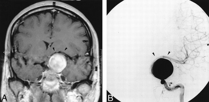
fig 1.
MR image and angiogram obtained prior to treatment.
A, Coronal T1-weighted MR image after gadolinium chelate infusion shows the giant left supraclinoid aneurysm shifting the left MCA (arrowheads).
B, Left ICA angiogram (frontal projection) depicts the left giant nonthrombosed supraclinoid aneurysm stretching the left MCA (arrowheads).

