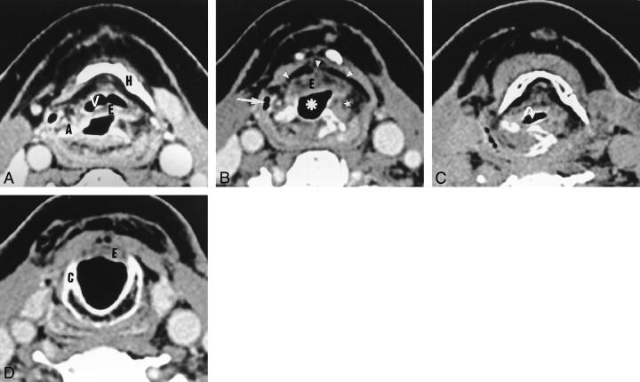
fig. 4. Axial CT scans obtained after SCPL with CHEP.
A, The vallecula (V) and epiglottis (E) are depicted at the level of the hyoid bone (H).
B, Below the level of the hyoid bone, the neovestibule (asterisk) is limited anteriorly by the epiglottis (E) and laterally by the NAFs (star). The lateral recess of the hypopharynx (arrow) and the preepiglottic space (arrowheads) are depicted.
C, During phonation, the neoglottis is oval and transversally oriented (arrow).
D, At the level of the cricoid cartilage (C), the subglottic lumen is empty and outlined by a thin mucosa. The inferior border of the epiglottis (E) cannot be distinguished.

