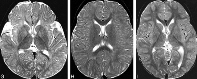
fig 4.
MR changes of myelination at the level of the basal ganglia.
A and B, Spin-echo (600/11 [TR/TE]) image (A) of a neonate shows that myelination (white matter with short T1 [hyperintensity on T1-weighted images] and short T2 [hypointensity on T2-weighted images]) is limited to the posterior limb of the internal capsule at this level. Spin-echo (3000/120) image (B) of the same patient shown in panel A.
C and D, Spin-echo (600/11) image (C) of a 5-month-old patient shows hyperintensity in the entire internal capsule, optic radiations, and splenium of the corpus callosum. Spin-echo (3000/120) image (D) of the same patient shown in panel C shows hypointensity limited to the posterior limb of the internal capsule and a portion of the optic radiations.
E and F, Spin-echo (600/11) image (E) of an 8-month-old patient shows hyperintensity in all white matter except the immediate subcortical regions. Spin-echo (3000/120) image (F) of the same patient shown in panel E shows hypointensity in the entire corpus callosum, the entire posterior limb of the internal capsule, and part of the anterior limb of the internal capsule.
G, Spin-echo (2500/80) image of a 12-month-old patient shows hypointensity in the entire internal capsule, in the subcortical white matter of the motor cortex, and in the subcortical white matter of the visual cortex.
H, Spin-echo (2500/80) image of an 18-month-old patient shows hypointensity in most of the deep white matter but lack of maturity of subcortical white matter.
I, Image of a 24-month-old patient shows that essentially all white matter is hypointense.

