fig 5.
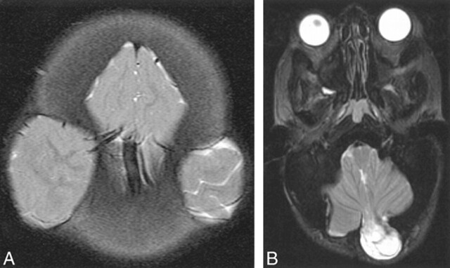
Acquired cephaloceles, surgical.
A, AROP. Axial T2-weighted FSE image. Bilateral decompressive craniectomies have resulted in a “Mickey Mouse”-like herniation of brain into and through the calvaria.
B, AROP. Axial T2-weighted FSE image. A posterior fossa decompressive craniectomy was complicated by a cephalocele. Cerebellar tissue, CSF, and meninges protrude through the surgical defect.

