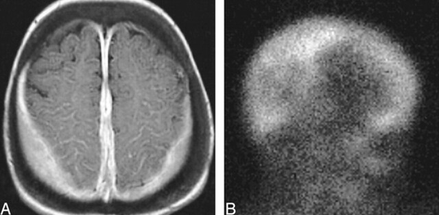
fig 8.
Extramedullary hematopoiesis, AROP.
A, Axial contrast-enhanced T1-weighted image shows homogeneously enhancing extraaxial tissue along the parietal convexities and falx cerebri.
B, Right lateral projection of 99mTc-sulfur colloid study shows uptake in these deposits of extramedullary hematopoiesis.

