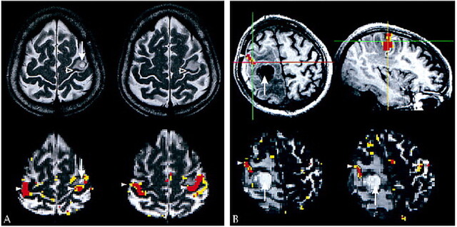
fig 1.
Adjacent to nonglial tumors, a normal volume of BOLD contrast enhancement was elicited.
A, Case 4. Arrows point to a small chronic abscess in the left precentral gyrus within the hand area. Upper row (T2-weighted turbo spin-echo sequence: 4500/120/1) shows edema apparently confined to the white matter. A narrow band of cortex is spared (open arrowheads). Lower row (echo-planar image: 3500/84/1), shows BOLD contrast enhancement in the cortex directly adjacent to the edema (arrowheads). Neither the edema nor the lesion affect the BOLD contrast enhancement.
B, Case 10. Arrows point to a metastatic lesion located in the right postcentral gyrus, slightly superior to the hand area. Arrowheads point to the functional activation of the sensorimotor cortex. Upper row is an overlay of the activation map onto the full head volume (MPRAGE: 9, 7/4/1 [TR/TE/excitations]). Lower row is an overlay onto the functional echo-planar sections (3500/84/1). In the ipsilateral hemisphere, the activation is squeezed between the swollen pre- and postcentral gyrus, but is not reduced. There is very little activation in the contralateral hemisphere.

