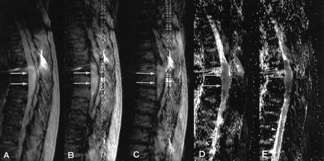
fig 2.
A–C, Diffusion-weighted images with a gradient b-values of 0 (A), 250 (B), and 1000 s/mm2 (C). On the diffusion-weighted image of higher b-value, tumor intensity remains high (arrow). The vertical artifact likely appeared because of pulsatile motion of superior sagittal sinus.
G–E, Calculated apparent diffusion coefficient maps. Apparent diffusion coefficient values of epidermoid cyst is 1.3 × 10⁻³ mm2/s (G) (arrow) and 1.6 × 10⁻³ mm2/s (arrowhead). The solid nature of the spinal epidermoid cyst is represented.

