
fig 1.
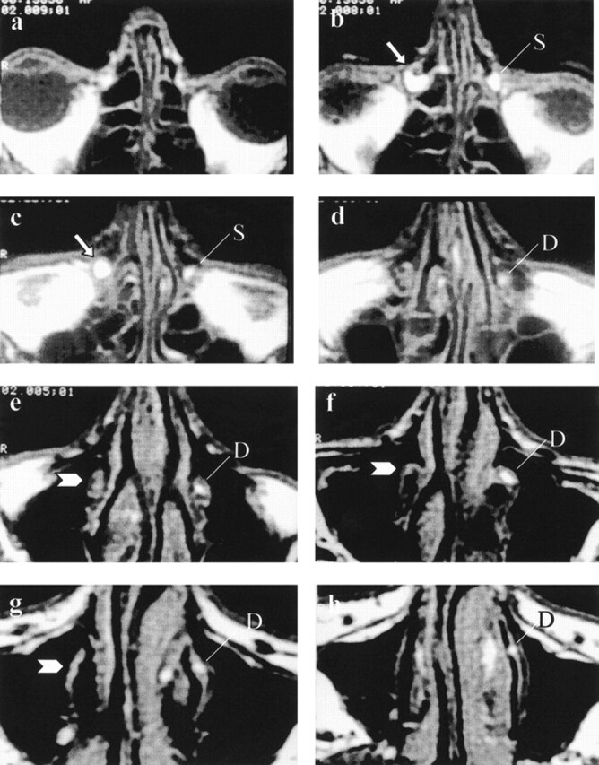
MRD images of a patient with right postlacrimal sac stenosis and healthy left lacrimal system. Axial T1-weighted (600/30/1 [TR/TE/excitations]) images obtained after the topical administration of diluted gadopentetate solution from the nasolacrimal duct to the orbital level. On the left side, healthy drainage of contrast-enhanced tear can be appreciated inside the lacrimal sac (L) and the nasolacrimal duct (N). On the right side, postsaccular stenosis causes dilation of the right lacrimal sac (arrows), and no contrast media is appreciated down into the ipsilateral nasolacrimal duct (arrowheads).
a, Nasolacrimal duct, most inferior section.
b through g, Progression of sections from inferior to superior.
h, Orbital level, superior section.