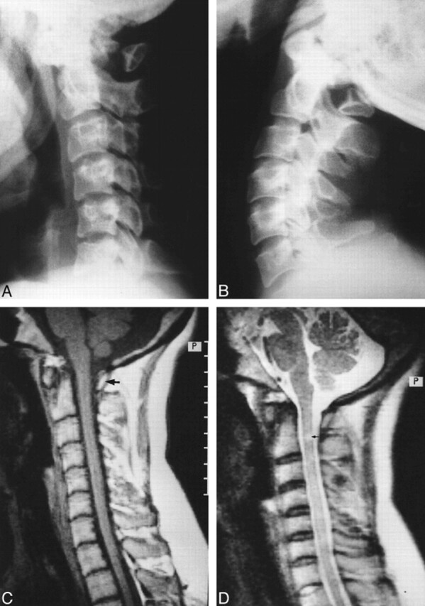
fig 1.

32-year-old woman with intermittent weakness and numbness of all four limbs.
A and B, Lateral radiographs of cervical spine taken in flexion (A) and extension (B) reveal partial aplasia of the posterior arch of the atlas with an isolated posterior bony fragment. Note the anterior displacement of the bony fragment during extension (B).
C, Midsagittal T1-weighted MR image through the cervical spine shows no compression of the cord by the posterior bony fragment (arrow).
D, Corresponding T2-weighted image shows a small, localized region of hyperintensity within the cord (arrow). Note that the signal alteration is seen slightly inferior to the anteroinferior margin of the isolated posterior arch remnant. A superimposed canal stenosis was noted from C2 through C5.
