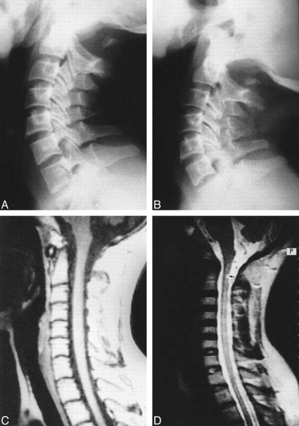
fig 2.

35-year-old woman with neck pain and paresthesias in both upper limbs.
A and B, In varying degrees of extension, plain radiographs show partial aplasia of the posterior arch of the atlas, with an isolated posterior tubercle. No obvious anterior displacement of the posterior tubercle is observed during extension.
C, Midsagittal T1-weighted MR image reveals no cord compression.
D, Corresponding T2-weighted image shows small, focal intramedullary hyperintensity (arrow) located just below the level of the posterior tubercle. In addition, there is focal canal stenosis at the C2–C3 disk level.
