fig 3.

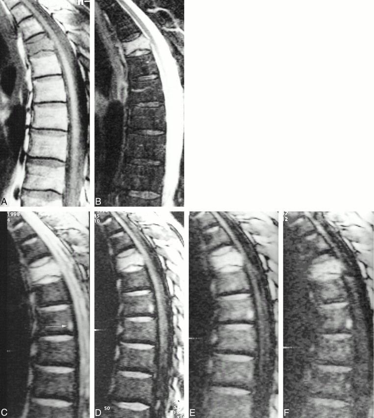
62-year-old man with a vertebral fracture due to a metastasis of a transitional cell carcinoma in the fifth thoracic vertebral body.
A, Hypointensity in the vertebra on T1-weighted SE images.
B, Corresponding STIR image (3600/60, TI = 150) shows homogeneous high signal intensity in the fractured vertebral body.
C–F, SSFP image of the same slice with increasing diffusion weighting (C = 0.6 ms, D = 3.0 ms, E = 6.0 ms, and F = 9.0 ms). The signal intensity is markedly hyperintense at δ = 0.6 to 6.0 ms and hyperintense at δ = 9.0 ms. There is a signal loss in the overall image with high diffusion weighting at δ = 9.0 ms. Note the focal area of hyperintensity in the seventh thoracic vertebral body posteriorly (arrow, C). This represents a fat island, because it is hyperintense on T1-weighted SE images and hypointense on STIR images. Fat has an extremely low diffusion coefficient and shows no signal loss on diffusion-weighted images.
