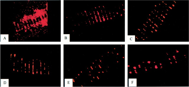
fig 10.
Segments of GDC coils co-cultured with fluorescence-labeled fibroblasts.
A, Fluorescent cells in culture adherent to the surface of the coil and extending into interstices of the coil lumen.
B, Immediately after withdrawal of coil into housing, the cells on the surface are stripped. Cells remain within coil lumen. Coils exposed to systemic arterial flow for 5 minutes (C), 10 minutes (D), 20 minutes (E), and 40 minutes (F). Numerous cells remain at all time intervals. The density of cells is diminished at the 20- and 40-minute time points relative to earlier samples.

