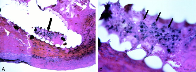
fig 5.
A, 100× H&E-stained 7-day sample. The arrow marks tissue that was present within the central lumen of the implanted coil. The basophilic, nucleated cells in the central region (arrow) are transplanted fibroblasts.
B, 400× magnification demonstrates fibroblasts (arrows) present within central lumen of coil.

