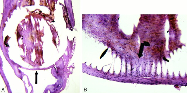
fig 6.
A, 40× H&E-stained view of 14-day speciment embolized with fibroblast-bearing GDC coil. The coil bridged the aneurysm neck, which developed neoendothelial lining.
B, 100× view of same specimen illustrates fibroblasts (arrow) present within the coil wind interstices and extending into the adjacent thrombus and aneurysm neck.

