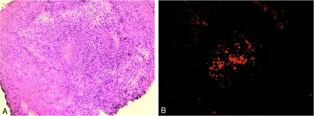
fig 8.
H&E (A) and fluorescent micrograph (B) of artery segment implanted 2 weeks previously with fluorescently labeled cells. The lumen of the arterial segment is nearly completely occluded, with a dense cellular infiltrate. The fluorescent images confirm the presence of labeled cells that are most intensely labeled in the central lumen of the vessel. Weak fluorescent labeling of the cells peripherally is likely secondary to the dilution effect on the membrane dye owing to cell division

