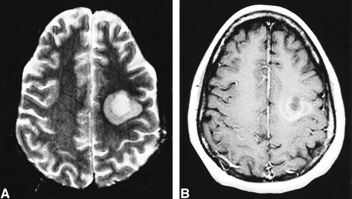
fig 1.

A, Fast spin-echo T2-weighted axial MR image through the centrum semiovale. This shows two separate concentric zones of demyelination in the left frontoparietal deep white matter represented as two different degrees of increased T2 signal (5300/82.2/1 [TR/TE/excitations]).
B, T1-weighted axial MR image after IV administration of Gd-DTPA at the same level as A. This clearly depicts multiple enhancing concentric rings representing two separate zones of active demyelination and increased blood brain–barrier permeability (550/9/1).
