fig 2.
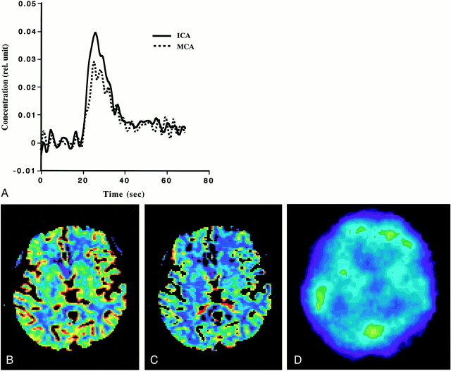
Example of PWI and 133Xe-SPECT studies in patient 7.
A, Comparison of the AIFs obtained from the ICA and MCA. Note that they vary slightly and that the AIF from the ICA is somewhat better than that from the MCA.
B, rCBF image generated from PWI (rCBF-PWI).
C, rCBV image generated from PWI (rCBV-PWI).
D, 133Xe-SPECT scan. Note that images generated from PWI were superior to 133Xe-SPECT scan in spatial resolution, and that rCBF and rCBV values in the deep brain structure can be evaluated on rCBF-PWI and rCBV-PWI images.

