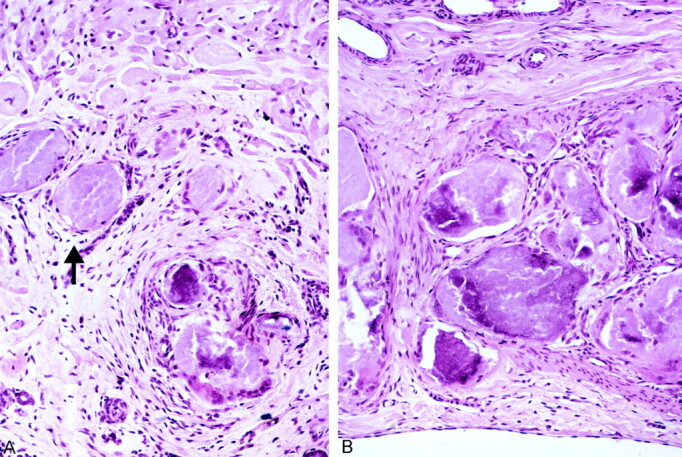
Fig 5.

Photomicrographs.
A, Three weeks after embolization, kidney shows hydroxyapatite microparticles (arrow) reaching the peripheral renal medullary arteries of approximately 200 μm in diameter. No arterial wall necrosis, extraluminal migration, or hemorrhagic changes are found. Several small round cells can be seen in the thrombus (hematoxylin and eosin; original magnification, ×100).
B, Histologic findings in larger arteries are nearly the same as those in smaller vessels (hematoxylin and eosin; original magnification, ×100).
