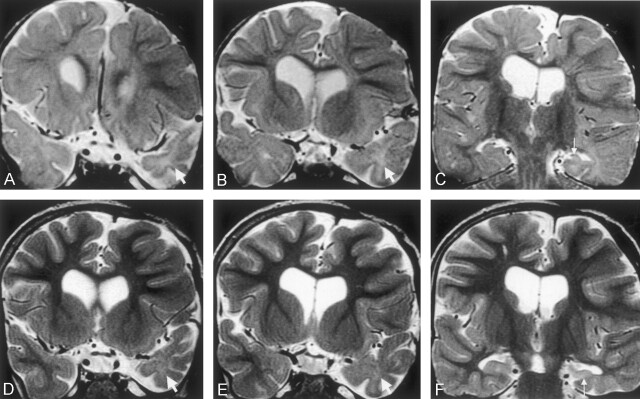
Fig 3.
Coronal view T2-weighted fast spin-echo images (2975/98) of a male patient who experienced onset of epilepsy at age 4 months, soon after drainage of a large left frontal subdural empyema. Clusters of complex partial seizures occurred every week during the first year of epilepsy.
A and B, Images obtained when the patient was 18 months old. An immature appearance persists in both temporal poles, but the ipsilateral left temporal pole is small (arrows), with abnormal increased white matter signal intensity.
C, Image obtained when the patient was 18 months old. Left hippocampal sclerosis with volume loss and increased signal intensity is shown (arrow). Note also the abnormal increased signal intensity in the temporal lobe white matter, compared with the delayed myelination seen elsewhere.
D and E, Images obtained at similar section positions when the patient was 5 years old. The myelination pattern in the frontal lobes and right temporal lobes is now mature. The left temporal pole is atrophic, and the ipsilateral left temporal white matter is now of similar signal intensity to gray matter, although still slightly hyperintense (arrows).
F, Image obtained when the patient was 5 years old. Typical left hippocampal sclerosis is shown (arrow), with a slight increase in white matter signal intensity on the left compared with the right on this more posterior section. Atrophy of the parahippocampal and fusiform gyri can be seen.

