Fig 1.
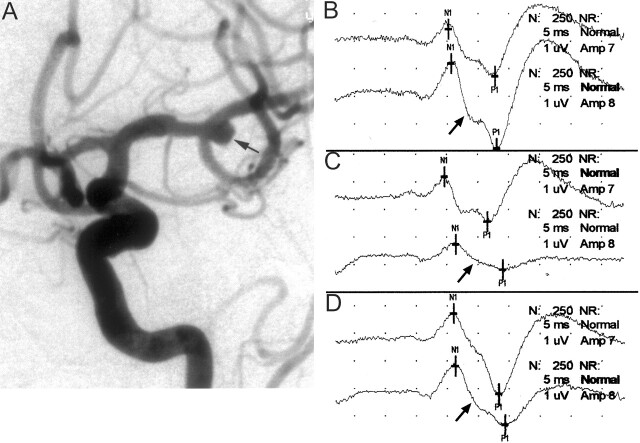
Images from the case of a 78-year-old woman who presented with symptoms of a subarachnoid hemorrhage.
A, Left internal carotid artery injection in the anterolateral projection shows a 5-mm left middle cerebral artery bifurcation aneurysm (arrow). Intra-aneurysmal coiling was attempted while the patient was systemically heparinized.
B, Baseline cerebral SSEPs after bilateral median nerve stimulation. Top tracing, left median nerve stimulation (ie, right brain); bottom tracing, right median nerve stimulation (ie, left brain) (arrow).
C, One minute after coil placement into the aneurysm, a >50% decrease in amplitude of the right median nerve SSEP was noted (arrow). This is consistent with significant left cerebral ischemia. Fluoroscopic evaluation suggested the coil was partially prolapsed into the parent artery, and considering the change in potentials, it was decided to quickly remove this coil. Formal angiographic assessment may well have shown significant compromise in the parent vessel; however, because the changes were rapid and profound, the coil was removed. Top tracing, left median nerve stimulation (ie, right brain); bottom tracing, right median nerve stimulation (ie, left brain) (arrow).
D, Left cerebral evoked potential (arrow) returned to baseline levels after removal of the coil. Because coil embolization could not be performed safely, the patient subsequently underwent surgical clipping of the aneurysm. Top tracing, left median nerve stimulation (ie, right brain); bottom tracing, right median nerve stimulation (ie, left brain) (arrow).

