Fig 2.
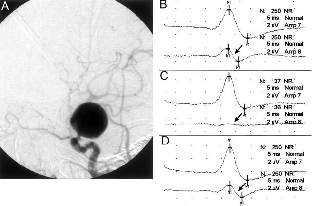
Images from the case of a 75-year-old woman who presented with a ruptured giant left internal carotid artery aneurysm.
A, Lateral projection angiogram of the left internal carotid artery shows the aneurysm arising in the supraclinoid segment shortly after the takeoff of the ophthalmic artery. The aneurysm was thought to be unfavorable for GDC embolization, and balloon test occlusion was thus performed in anticipation of permanent vessel occlusion. Because of the patient’s physical condition, the procedure could be performed only with the patient under general anesthesia.
B, Baseline cerebral SSEPs after bilateral median nerve stimulation. Note the baseline asymmetry, with the right median nerve SSEP (arrow) being smaller in amplitude than the left. Top tracing, left median nerve stimulation (ie, right brain); bottom tracing, right median nerve stimulation (ie, left brain) (arrow).
C, Left internal carotid artery balloon test occlusion was performed, resulting in gradual amplitude reduction of the left cerebral (right median nerve stimulation) SSEP over 5 min and precipitous decrease in the 6th min. The SSEP obtained 6 min after balloon occlusion shows a nearly complete loss of the left cerebral SSEP (arrow). Top tracing, left median nerve stimulation (ie, right brain); bottom tracing, right median nerve stimulation (ie, left brain) (arrow).
D, Balloon was deflated immediately after the SSEP tracing shown in 2C. The cerebral SSEP returned to baseline amplitude levels after 1 min (arrow). Top tracing, left median nerve stimulation (ie, right brain); bottom tracing, right median nerve stimulation (ie, left brain) (arrow).

