
fig 1.
Case 1.
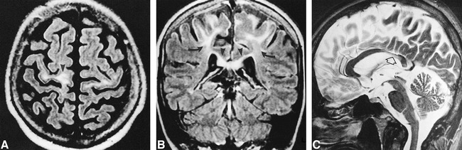
A, Axial FLAIR image (9264/150/2; TI = 2200) shows increased signal intensity of the white matter of the precentral gyri, which are atrophic (arrowhead).
B, Coronal FLAIR image (9264/150/2; TI = 2200) shows the hyperintensity limited to the upper part of the motor cortex (arrows) and extending across the corpus callosum (arrowheads).
C, Midline sagittal turbo spin-echo T2-weighted image (5422/110/2) shows thin, slightly hyperintense isthmus of the corpus callosum (arrow).