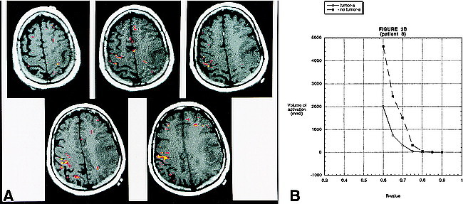
fig 2.

A, BOLD fMR activation from a finger-tapping paradigm coregistered to an axial T1-weighted image (500/14 [TR/TE]) in patient 8. Pathologic analysis revealed a left frontoparietal glioblastoma multiforme. The areas in yellow correspond to an R value of 0.72. The areas in red correspond to an R value of 0.67. There is robust activation in the right motor cortex. It is interesting to note that the activation is seen along the most anterior and posterior aspects of the precentral gyrus. This is the location of the cortical gray matter. The central portion of the gyrus (which contains the white matter tracts) does not reveal activation. On the right side, activation is appreciated in the postcentral gyrus, which most likely represents the sensory cortex. Activation is also identified in the left motor cortex; however, the volume is much less than on the side without the tumor. The activation on the left side is seen in the most superior and medial part of the precentral gyrus—the area relatively spared by the glioma.
B, A graph of the volume of activation for different R values for patient 8. As in figure 1, for all R values that reveal activation, the volume of activation is greater on the side opposite the tumor. The absolute volume of activation and the pattern of activation, however, differ from patient 9.
