fig 3.

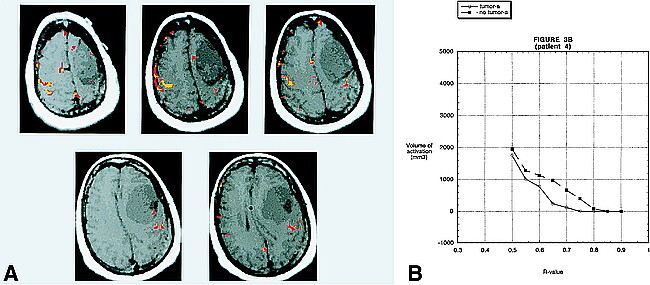
A, BOLD fMR activation from a finger-tapping paradigm coregistered to an axial T1-weighted image (500/14 [TR/TE]) in patient 4. Pathologic analysis revealed a left frontal lobe meningioma. There is prominent activation of the motor cortex on the side contralateral to the tumor, at R values of 0.70 (yellow) that allows for accurate delineation of the primary motor cortex. At these R values, there is very little activation on the side with the tumor. At R values of 0.55 (red), however, the volume of activation in the primary motor cortex of the side with the tumor approaches the activation on the contralateral side. This occurs before the onset of “noise” in areas of the brain not related to the neural control of motor function. This example emphasizes the need to evaluate each patient individually at multiple R values. Areas of activation located outside the brain over the right frontal convexity probably represent cortical venous drainage.
B, A graph of the volume of activation for different R values for patient 4. There is a large difference in the volume of activation between the two sides for R values between 0.65 and 0.75. At 0.50, however, the volume of activation on the two sides is almost the same.
