
fig 2.
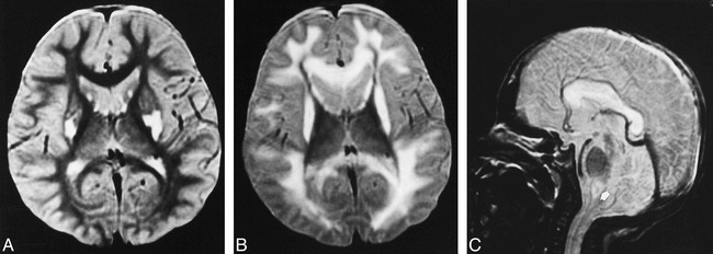
Patient 2: group BG lesion pattern.
A, Axial T2-weighted image (2000/80/2) at the age of 3 years shows increased signal intensity in the bilateral putamina and caudate heads, but no detectable brain stem lesion. Clinical findings included mental retardation, muscle weakness, and failure to thrive.
B and C, Axial (B) and sagittal (C) T2-weighted images (2000/80/2) at the age of 9 years show static and atrophic lesions in the bilateral putamina and progressive lesions in the cerebral white matter, corpus callosum, and medial medulla oblongata (arrow, C). Clinical findings included irregular breathing, lethargy, and inability to feed.