fig 5.

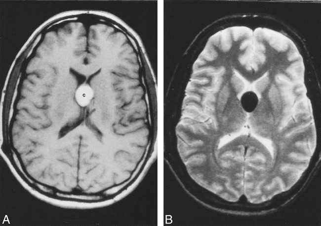
In vivo MR imaging of a colloid cyst (different patient).
A, Axial noncontrast T1-weighted image shows oval-shaped, hyperintense colloid cyst [c].
B, Corresponding T2-weighted image shows the cyst to be markedly hypointense. There is no hydrocephalus in this patient.
