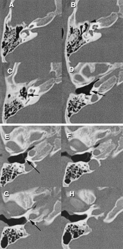
fig 1.

Axial Images (A–H), from superior to inferior. The small vascular channel can be seen leaving the vertical carotid at G (arrow), and can be followed along the lower medial wall of the middle ear (D–F) (arrows). Level C is at the plane of the stapedial crura. The vessel cannot be clearly separated from the anterior crus of the stapes. After following the facial nerve canal, the small vascular channel reaches the middle cranial fossa at level B (arrowhead). Level A represents the position of the geniculate ganglion turn of the facial nerve canal
