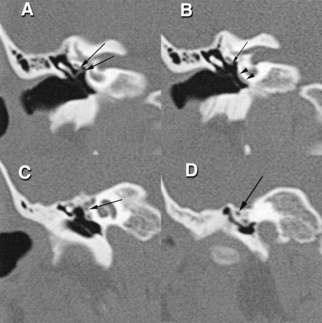
fig 2.

Coronal images (A–D), from posterior to anterior. The narrow presumed vascular structure is seen at B (arrowheads), coursing along the promontory. At level A, the small vascular structure (arrows) crosses the oval window niche, to end in the lower inferomedial aspect of the tympanic segment of the facial nerve canal. This small channel is also seen at B (arrow). More anteriorly (C and D), the small canal (arrows) courses just inferior to the tympanic segment of the facial nerve canal in a separate channel as it passes toward the floor of the middle cranial fossa. The small soft-tissue structure immediately inferior to this canal is the tensor tympani muscle within its semicanal
