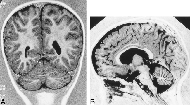
fig 7.

Patient 17. MR imaging findings in a 1 year-old boy with facial deformities, hypotonia, and developmental delay.
A, Coronal IR T1-weighted image (11520/60/400/2) shows medial vermian fissure (arrow).
B, Sagittal IR T1-weighted image (same parameters) shows lack of normal fissures of the vermis associated with inferior vermis hypoplasia.
