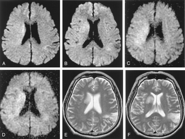
Fig 3.
A–F, A 64-year-old man who had left hemiparesis for 1 hour 20 minutes. Initial b = 1000 images reveal subtle hyperintensity in the right corona radiata and basal ganglia (A, B). The lesion is more conspicuous and extensive on b = 2000 images (C, D). Follow-up T2-weighted image obtained 3 days later shows progression to infarction at the basal ganglia (E, F). Initial diffusion change is partly normalized in the posterior portion of the initial lesion. An old infarction is noted in the left posterior temporal cortex on the T2-weighted image.

