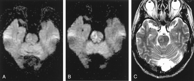
Fig 4.
A–C, A 57-year-old man who had left hemparesis for 4 hours 40 minutes. Initial b = 1000 image reveals subtle hyperintensity in the right pons (A). The lesion is more conspicuous on the b = 2000 image (B). Follow-up T2-weighted image obtained 2 days later shows the infarction at the corresponding location.

